Continuum of Care
The Future of Breast Health
Explore The Continuum
Products
Request Information
Continuum of Care
The Future of Breast Health
Explore The Continuum
Products
Request Information
This is my archive
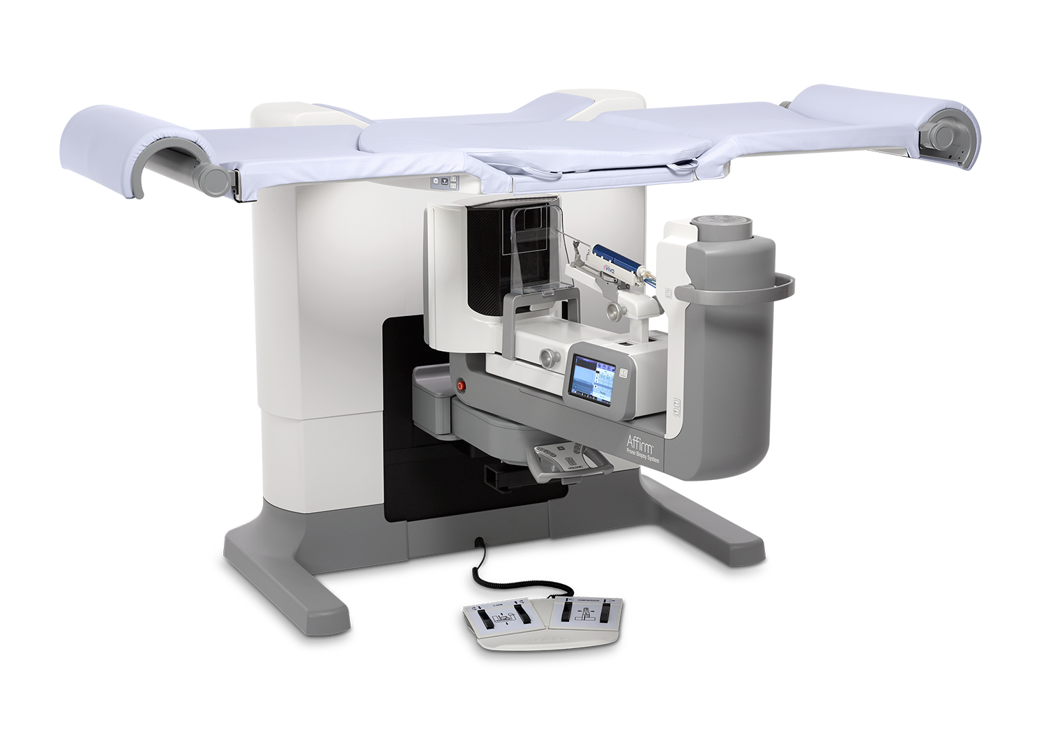
Affirm
®
Prone Biopsy System
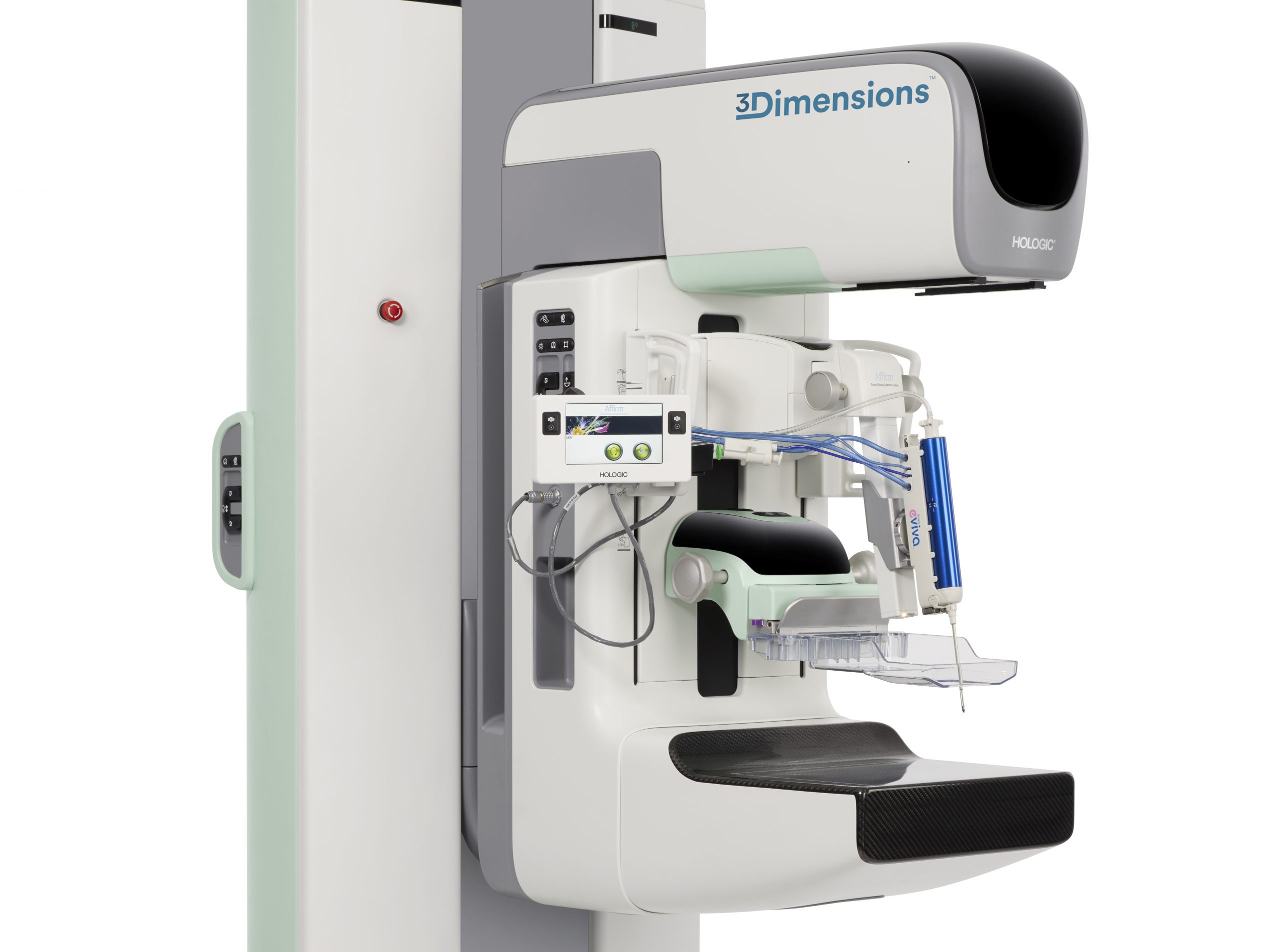
Affirm
®
Upright Breast Biopsy Guidance System